
Analysis of cases with deep coverage and deep curve
- Age:14
- Gender:male
- Chief complaint: Mouth protrusion, tooth protrusion
- Use product:PT 5 Plus
- Treatment cycle:19 months
Basic information of patients
Gender: MaleMedical history: None
Dental treatment history: None
Complaint: Mouth protrusion, tooth protrusion
Previous History: None
Correction start date: July 17, 2021

Inspection&Diagnosis
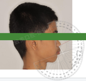
1、Facial analysis
Facial photo
Positive analysis

Side analysis

Face type: ovoid
Symmetry: facial asymmetry
Chin: slightly left deviation
Pupil connection: parallel
Angular connection: parallel
Facial ratio: 1/3 of the lower face is slightly longer, and the thickness of the upper and lower lips is thicker
Face protrusion: Raised face type
Upper lip protrusion: protrusion
Lower lip protrusion: protrusion
Chin protrusion: later retracted
Chin neck angle: right angle
Nose chin lip angle
Nasolabial angle: 93 °
(Reference range: 90 ° -95 °)
Chin lip angle: 103 °
(Reference range: 120 ° -130 °)
Vertical line of the ideal anterior boundary of the upper jaw
Determine the target position in the natural head position;
The distance between the measured target and the initial FA point is 5mm.
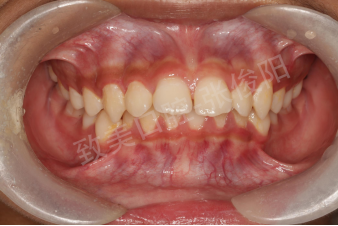
Lip tooth relationship
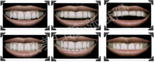
Laughing line: high position
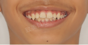
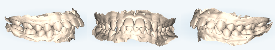
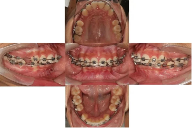
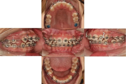
Intraoral examination
Intraoral irradiation
1. The maxillary gap is 2mm, and the mandibular crowding is 3mm;
2. Local redness and swelling of the gums.
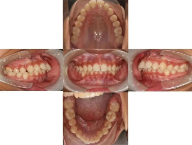
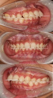
Horizontal relationship
The upper and lower dental arches are basically symmetrical

Vertical relationship
1. Second degree deep overlap of anterior teeth;
2. The speed curve depth is 3.5mm.
Forward backward relationship
1. Lip inclination of upper and lower anterior teeth;
2. Front teeth covered with 5mm;
3. The relationship between the right molar is neutral to the middle, and the relationship between the right canine and the far middle is pointed to the apex;
4. The relationship between the left molar is neutral, and the relationship between the left canine and the distal apex is pointed to the apex.

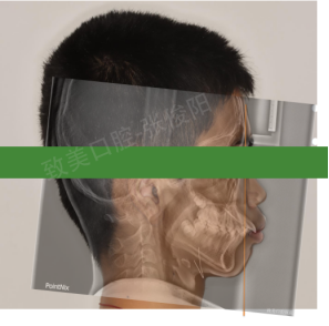
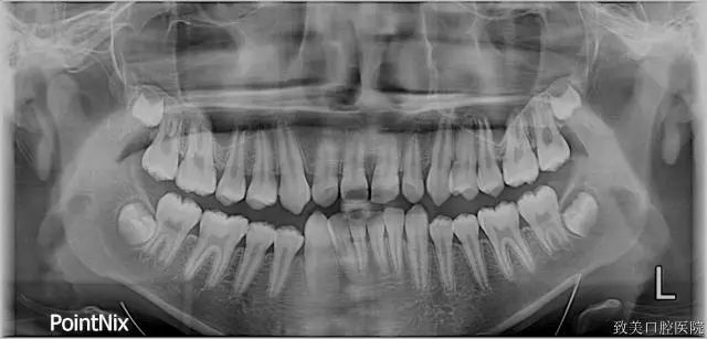
3、X-ray examination
Panoramic film
1. Teeth: 18, 28, 38 tooth germs present, root length is normal, and periodontal ligament is normal;
2. Joint: Slightly asymmetric joint shape without obvious tenderness;
3. Asymmetric lifting height;
4. The maxillary sinus is slightly lower.
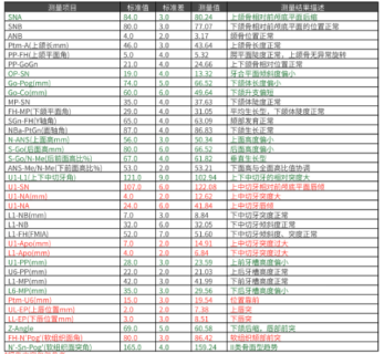
lateral cephalogram
Normal airway
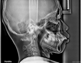
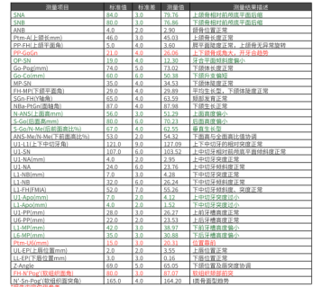
Analysis of cephalometric measurement results:
4、Model Check
1. Molar relationship: The relationship between the right molar is neutral to the middle, and the relationship between the left molar is neutral;
2. Canine relationship: the right canine relationship is distal to apical, and the left canine relationship is distal to apical;
3. Crowding degree: 2mm scattered gaps in the upper jaw and 3mm crowded in the lower jaw;
4. Center line: the upper center line is aligned, and the lower center line is 1.5mm to the right;
5. Bolton: 78.9% for anterior teeth and 91.9% for complete teeth.
5、Problems and Diagnosis
problem
1. Smile with exposed gums, with the smile line facing away from the tangent;
2. Lip inclination of upper and lower anterior teeth;
3. Deep covering and deep covering;
4. Uneven dentition;
5. 2mm to the right of the lower centerline
6. The speed curve is steep.
diagnosis
1. An's Class I;
2. Uneven dentition.
6、Corrective goals and plans
Corrective goals
1. Improve the convex shape;
2. Align and level the upper and lower dentition;
3. Open the bite and establish normal coverage of the anterior teeth;
4. Establish a neutral relationship between molars and canines;
5. Align the upper and lower center lines with the center line of the surface.
Correction plan
1. Remove four 4;
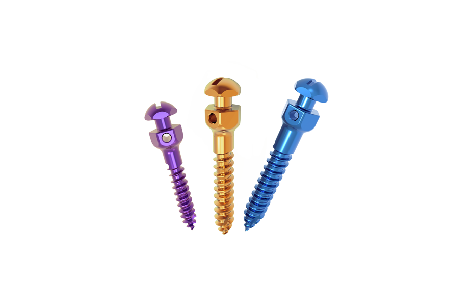
2. Implantation of anchorage nails in maxillary posterior teeth;
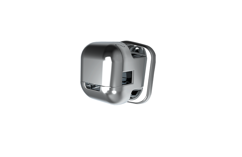
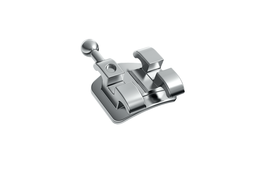
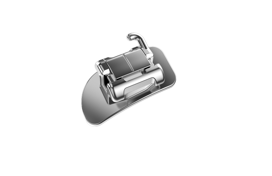
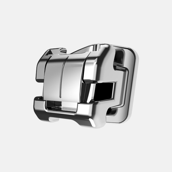
3. Use Pute PT 5 Plus self-locking bracket - standard torque to align and level the upper and lower teeth;
4. Close the extraction gap by retracting the anterior teeth;
5. Establish normal coverage of anterior teeth, with a neutral relationship between molars and canines.
Treatment process
September 25, 2021
Replace the upper jaw with 0.017x0.025nt, and lower the lower anterior teeth with 0.018nt to lower the auxiliary arch.
November 20, 2021
Implantation of anchorage screws in the posterior teeth of the upper jaw, replacement of 0.017x0.025ss anterior teeth in the upper jaw, and replacement of 0.016x0.025nt in the lower jaw.
March 13, 202
Close the gap between the upper and lower retracted anterior teeth
June 13, 2022
Apply positive torque to the anterior teeth of the upper jaw, and continue to retract upwards and downwards.

February 4, 2023
End orthodontic treatment
After treatment
1、Comparison of panoramic films
2、Comparison of lateral cephalometric films

3、Cephalometric overlap map

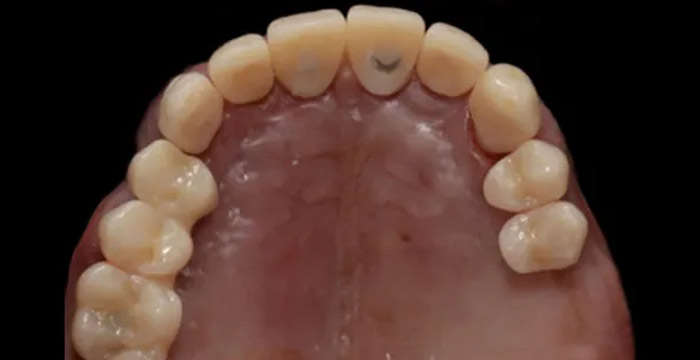
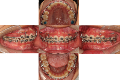
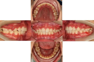
4、Intraoral contrast
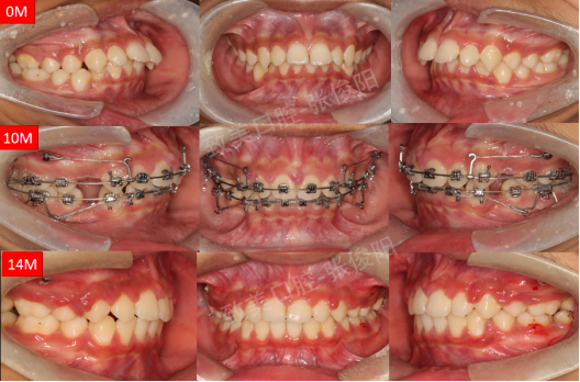
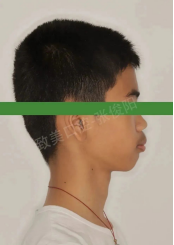
5、Facial contrast
Face to face comparison

Side comparison

Summary and Experience Sharing
1. In the early stage, open the bite by lowering the auxiliary arch and flattening the curve of the lower anterior teeth;
2. High position nails combined with long traction hooks prevent the side effects of adduction clock swing and lower the upper anterior teeth;
3. Strengthen the strong anchorage of the upper jaw and retract the upper anterior teeth;
4. Three types of traction were used during the surgery to reverse the rotation of the jaw plane and improve the morphology of the chin;
5. Patients with thick lips require long-term lip muscle training to improve their mouth shape and lateral appearance.
RELATED NEWS
-

-
-
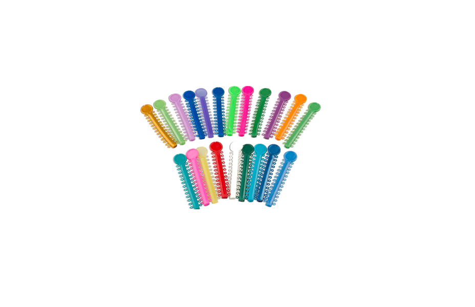
Tips On Choosing and Using Orthodontic Rubber Chainsnews | 2023-12-18
We are at your service.
Whether you have inquiries about our products or need assistance with troubleshooting, our team of sales and service experts is here to assist you.
GET A QUOTE